Real-time sonoelastography and ultrasound evaluation of the Achilles tendon in patients with diabetes with or without foot ulcers: a cross sectional study
Evranos B, Idilman I, Ipek A, Polat SB, Cakir B, Ersoy R
J Diabetes Complications. 2015 Aug 20. [Epub ahead of print]
Introduction
당뇨는 대사이상과 장기간 합병증으로 특징지어지는 내분비장애이다. 단백질의 non-enzymatic glycation은 당뇨에서 합병증 발생에 중요한 역할을 한다. 아킬레스건(AT)은 인체에서 가장 강하고 크며 두꺼운 힘줄이고 발의 생체역학에 중요한 역할을 한다.
AT는 기계적인 충격을 흡수하고 propulsion시에 longitudinal arch를 안정시키고 착지기간에는 붕괴되는 것을 방지한다. AT의 static stiffness가 전족부 부하를 증가시키는데 중요한 역할을 하는 것으로 보고되고 있는데 이로 인해 족부궤양을 발생시키는 병리학적 과정이 시작되는 것으로 생각되고 있다. 고혈당에 기인한 nonenzymatic glycation에 의해 근육, 연골, 힘줄 및 인대를 포함하는 대부분의 인체 조직에 당화 최종산물이 축적된다. AT 두께는 제1형 및 제2형 당뇨 환자에서 증가되는데 특히 다발성 신경병증이 있는 환자에서 더욱 그렇다.
Sonoelastography (SE)는 다양한 조직과 병변의 탄성과 강직에 대한 정보를 제공하는 새로운 초음파영상기법이다. SE는 다양한 임상영역에 적용되고 있으나 근골격계 질환과 당뇨환자들에서는 아직 적극적으로 사용되지 않고 있다. 실시간 SE는 힘줄의 경도에 대한 유용한 정보를 제공하고 B-mode 초음파로 보완할 수 있다. SE의 color-coded image에서 정상 힘줄은 단단하기 때문에 파란색과 녹색 음영으로 보이고 병적으로 변성되면 힘줄의 강직이 감소하게 되어 중등도 연화된(softening) 경우는 노란색으로 중증인 경우에는 빨간색 음영으로 보인다.
이에 저자들은 발 질환이 있거나 없는 제2형 당뇨 환자들의 AT 두께와 강직을 평가하고 영향을 미치는 요인들을 연구해보고자 하였다.
Material and Methods
78명의 당뇨환자 중 발 궤양이 있는 35명의 환자를 group I으로 하였고 발 궤양이 없는 43명의 환자를 group II로 하였으며 연령, 성, BMI를 고려한 건강한 대상자 33명을 대조군으로 하였다. 모든 대상자에게 AT의 초음파검사와 SE를 시행하였으며 아킬레스건 두께(ATT), 강직(ATS)를 측정하였다. 또한 모든 당뇨 환자들은 fasting plasma glucose (FPG) 와 glycosylated hemoglobin (HbA1C) 검사를 받았으며 그 외의 당뇨 합병증과 관련된 검사들을 받았다.
각각의 AT는 axial과 longitudinal plane에서 검사하였으며 AT는 근위부 1/3(근건 접합부), 중간 1/3(종골 부착부로부터 2-6 cm), 원위부 1/3(종골 부착부)로 구분하였다. 각 세 부위의 전후방 거리는 transverse scan으로 측정하였고 실시간 SE 영상은 longitudinal plane에서 확인하였다. 조직 탄성 분포는 실시간으로 측정하였고 결과는 B-mode 영상으로 겹쳐진 color map으로 표시되었다. 색 범위는 파란색(hard)부터 빨간색(soft) 까지 이었고 조직의 상대적인 강직 정도를 나타내었다.
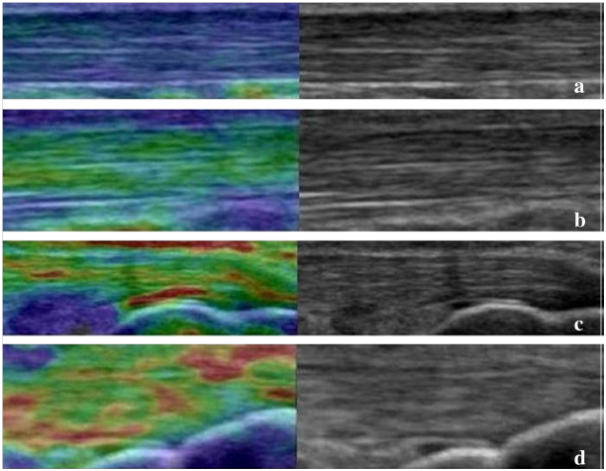
힘줄의 탄성은 두 가지의 주된 형태, 즉 type 1은 파란색/녹색 (hard tissue), type 2는 녹색 내의 노란색/빨간색 (intermediate-soft tissue)으로 구분하였다. 이들 type을 다시 두 subtype으로 정의하였다. Type 1a blue predominance, type 1b green predominance, type 2a small yellow/red areas within green predominance, type 2b small green areas within yellow-red predominance. 이 정의에 따르면 type 1a에서 2b로 갈수록 조직의 탄성은 단단함에서 부드러움으로 진행된다.

Fig. 1. The elasticity of tendons by sonoelastography. (a) Type 1a blue predominance; (b) type 1b green predominance; (c) type 2a small yellow/red areas within green predominance; (d) type 2b small green areas within yellow–red predominance. The elasticity of the tissue was in a spectrum ranging from hard to soft as the type progresses from 1a to 2b.
Results
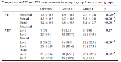
AT는 Group II와 대조군에 비해 Group I이 의미있게 두꺼웠다. AbA1C, FPG와 당뇨유병기간은 Group I이 높았다. ATT는 Group I에서 신경병증, 망막병증, 신장병증, 말초동맥질환, 관상동맥질환과 양의 상관관계를 보였으나 Group II에서는 상관관계를 볼 수 없었다. ATS는 Group II와 대조군에 비해 Group I에서 감소되어 있었다.

Conclusion
당뇨환자에서 AT 구조의 변화가 발질환에 선행되는 것으로 생각된다. 이 연구는 당뇨환자에서 AT의 SE 결과와 ATT와 다른 당뇨합병증과의 상관관계를 처음으로 보고한 논문이다.


